Biocinematics

Illustration

Osteocalcin and Bone Mineral

IllustrationStuart JantzenComment

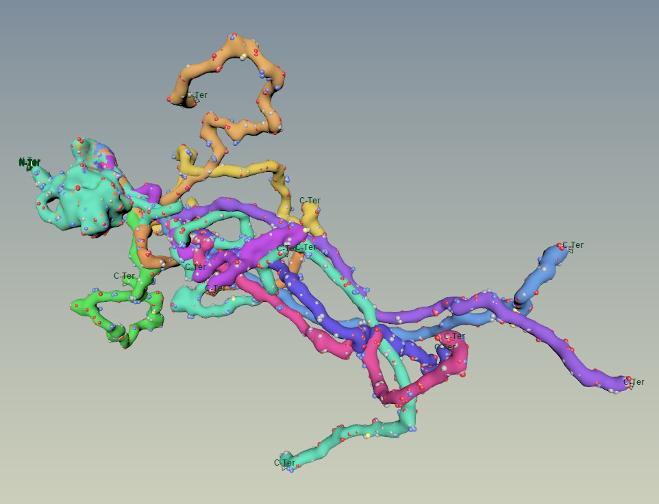
April's Molecule of the Month at the Protein Data Bank features proteins that bind to biominerals. This is osteocalcin binding to calcium ions, a major component of the calcium-phosphate lattice that makes up hydroxyapatite. Hydroxyapatite is essentially "bone mineral", which helps gives bones strength and rigidity.

The glowing spheres are the calcium ions that osteocalcin is currently binding.

The glowing spheres are the calcium ions that osteocalcin is currently binding.

This illustration was the first one where I brought molecular data directly into Houdini, the new 3D application that I have been learning over the past few months. Houdini requires very different workflows from other 3D applications, so learning how to manipulate data for this illustration was interesting and insightful, and I believe processes like this will allow me to create some pretty interesting content in the future.

The green balls here are the important calcium ions. It didn’t take too many nodes to generate this, which is nice.

The green balls here are the important calcium ions. It didn’t take too many nodes to generate this, which is nice.

Houdini can natively read PDB data files like the one I downloaded for osteocalcin, however beyond some helpful data organization, it doesn't have any tools for displaying different molecular representations, so everything from space-filling representations (like the one above) to surface meshes to backbones must be created from scratch. Fortunately Houdini is a great tool for building tools, so I'll be spending some time developing an internal toolkit to show molecules in different visual styles.

A single hydroxyapatite unit: Ca in green, PO4 in yellow/red, and OH in red/white

A single hydroxyapatite unit: Ca in green, PO4 in yellow/red, and OH in red/white

The crystal data for the bone mineral was in a different format (CIF), so I used UCSF Chimera to export a new PDB file and get the measurements that allowed me to expand a single atomic "cell" to the full crystal field of repeated units.


Thanks for reading,

Stuart

Measles Virus Nucleocapsid

IllustrationStuart JantzenComment

In continuing my illustration series following the Molecule of the Month over at the Protein Data Bank (1), this month I created an image showing the nucleocapsid of the measles virus.

Measles Virus Nucleocapsid - March Molecule of the Month

Measles Virus Nucleocapsid - March Molecule of the Month

I'm sure you've heard of measles before. In fact, it seems to be a bit of an ongoing newsworthy item now and again. Measles is caused by a virus that is extremely contagious and can be deadly (2). Fortunately, we have a widely available vaccine (typically administered together with protection against mumps, rubella, and sometimes varicella (3)) to prevent the spread of this illness.

This image doesn't show the entire virus, instead it focuses on the genetic material (RNA in red) and the protein coat (grey) that protects the viral genes from our bodies' natural defenses (4) and also plays a role in helping the virus make copies of itself through transcription and RNA replication (5).

Making of

I spent a long time looking at the proteins amazingly illustrated by David Goodsell on the RCSB website, and wondering how I might create an illustration of my own. I liked the idea of the long repetitive pattern of the nucleocapsid. When I did some more research, I realized that a long flexible tail was omitted from the structural data (4) and the close-up illustration of the nucleocapsid. Working with my recent method (explained in this YouTube tutorial) I decided to append the flexible tail to the existing data and create something (hopefully) striking. Although the tail originates toward the hollow space in the center of the complex, there is evidence that the tail feeds itself back toward the outside (there's also space limitations with stuffing it all inside), resulting in an external coat of long flexible fibers (6,7).

PDB 4UFT

PDB 4UFT

Once I had that piece in place, it became clear that I could create something quite menacing, using visual inspiration from the Sentinels in The Matrix (which is 20 years old this month!) (8). This approach did have some technical and visual challenges to work out.

A bunch of simulations all stacked up

A bunch of simulations all stacked up

Instead of making 2,516 copies (9) of the nucleoprotein and simulating all of them individually, I decided to simulate a lower number of copies and create static meshes that could be randomly scattered along the length of the nucleocapsid. I figured out the correct transformation offsets I would need to spiral the copies correctly, and used MASH to create a single unit of 37 proteins. Side note: Yes, although I'm learning Houdini, I did use Maya for this illustration because I already know the tools well and I have yet to figure out a pipeline for getting molecular data directly into Houdini.

37 proteins and 222 RNA nucleotides

37 proteins and 222 RNA nucleotides

Then I adjusted the random seed to make several more 37-monomer "units", and sent them over to ZBrush for some retoplogy to reduce the mesh density for all the background units. I also made a lower-resolution RNA spiral unit.

Tip: After retopology of thin meshes, use the 3D Gizmo and ctrl+drag the yellow center to inflate the model closer to the original volume.

Tip: After retopology of thin meshes, use the 3D Gizmo and ctrl+drag the yellow center to inflate the model closer to the original volume.

Turns out the RNA is all cytosine residues… so not perfectly accurate… shhh!

Turns out the RNA is all cytosine residues… so not perfectly accurate… shhh!

 Finally, I used MASH again to spread the units along a curve, and posed the whole assembly to my liking.

I believe this is approximately the correct full length of the measles genome, and yes it should fit inside a typical measles capsid. Successful assembly!

I believe this is approximately the correct full length of the measles genome, and yes it should fit inside a typical measles capsid. Successful assembly!

I feel like the RNA is the most central and dangerous aspect of this virus, so I wanted all of the color in the image to come from that and have it draw the eye. Because of how the protein binds to the RNA, I didn't get as clean a view of the RNA helix as I was hoping, but I think overall the image works as intended. I hope you like it.

No connection to the Xbox Red Ring of Death

No connection to the Xbox Red Ring of Death

Thanks for reading,

Stuart


References (fancy!):

  1.  PDB101: Molecule of the Month: Measles Virus Proteins. RCSB: PDB-101 Available at: http://pdb101.rcsb.org/motm/231. (Accessed: 12th March 2019)

  2. Measles. World Health Organization (2018). Available at: https://www.who.int/news-room/fact-sheets/detail/measles. (Accessed: 15th March 2019)

  3. MMR Vaccination | What You Should Know | Measles, Mumps, Rubella | CDC. Centers for Disease Control and Prevention (2018). Available at: https://www.cdc.gov/vaccines/vpd/mmr/public/index.html. (Accessed: 15th March 2019)

  4. Gutsche, I. et al. Near-atomic cryo-EM structure of the helical measles virus nucleocapsid. Science 348, 704–707 (2015).

  5. Jiang, Y., Qin, Y. & Chen, M. Host–Pathogen Interactions in Measles Virus Replication and Anti-Viral Immunity. Viruses 8, (2016).

  6. Jensen, M. R. et al. Intrinsic disorder in measles virus nucleocapsids. Proc. Natl. Acad. Sci. U. S. A. 108, 9839–9844 (2011).

  7. Desfosses, A., Goret, G., Farias Estrozi, L., Ruigrok, R. W. H. & Gutsche, I. Nucleoprotein-RNA Orientation in the Measles Virus Nucleocapsid by Three-Dimensional Electron Microscopy. J. Virol. 85, 1391–1395 (2011).

  8. The Wachowski Brothers. The Matrix. (1999).

  9. Lund, G. A., Tyrrell, D. L., Bradley, R. D. & Scraba, D. G. The molecular length of measles virus RNA and the structural organization of measles nucleocapsids. J. Gen. Virol. 65 ( Pt 9), 1535–1542 (1984).

A Fresh Start

Misc, IllustrationStuart JantzenComment

If you've been following my work for any length of time, you're probably aware that I've kept a blog over at biocinematics.blogspot.com. This week I reached the 10th anniversary of that blog and decided it was a good time to "reboot" the blog fresh and new on this site.

 So here we are!

…..

..

.

Blank Page Syndrome!

.

.

Oh, I'll share the first image from a series of a molecular illustrations I started which is inspired by the Molecule of the Month series created by David Goodsell at the RCSB Protein Data Bank.

Initiation_Factor_eIF4E

The February Molecule of the Month is the eukaryotic initiation factor eIF4E. By binding to the 5' cap of mRNA (seen in the lower part of the image), eIF4E helps bring mRNA and the ribosome together to start protein synthesis. eIF4E also features a disordered flexible "tail" (in the upper left) which is involved in binding other parts of the initiation complex.

If you are on Instagram, I suggest following me @biocinematics to see more of this series as I create more molecular portraits.

On a related note, if you haven't yet seen anything on my "Blog 1.0", and you want to catch up a bit on recent news, I'd encourage you to have a peek at these posts:

10th Anniversary: The end of one blog and the beginning of another (and my demo reel!)

Knots and Robots: Recent client work

And now for something (almost) completely different: Job changes and new beginnings


If you want to read some humorous off-topic stuff, I'd recommend these:

Changing a Hard Drive

Bad auto transcription

One thing I would strongly caution against is going right back to 2009 to see the ugly beginnings of learning difficult software. Or… /shrug I can't stop you.


Thanks for reading!

Stuart